Cornea Diseases & Cornea Transplantation Surgery
|
|
Keratoconus 2020 & 2021 videos above and See her work on corneal research here.
Keratoconus
What is Keratoconus?
The cornea is the clear transparent window at the front of your eye in the shape of a round dome. If you wear contact lenses, the cornea is where the contact lens rests on. It is responsible for accurately focusing light into the back of the eye. In keratoconus, the cornea thins out and becomes uneven, resulting in an irregular cone shaped cornea. As a result, the light rays entering the eye become out of focus and your vision becomes blurry and distorted.
What are the symptoms of Keratoconus? How is keratoconus treated? What is Cornea Cross Linking (CXL)?
Keratoconus starts in your teens. Early symptoms are worsening vision that require regular change in glasses with increasing astigmatism. Once keratoconus progresses to the late stages, your vision will be poor and may not be correctable with glasses or contact lenses. When this happens a cornea transplant is required (CAIRS, DALK or PK). Cornea Cross Linking (CXL) is a new treatment which can halt or slow the progression of keratoconus. This aims to prevent the late stages of disease. However this is only effective in the stage of early progressive disease. This is why early diagnosis and monitoring in keratoconus is important.
How is Keratoconus diagnosed? How do I know if I have early progressive disease?
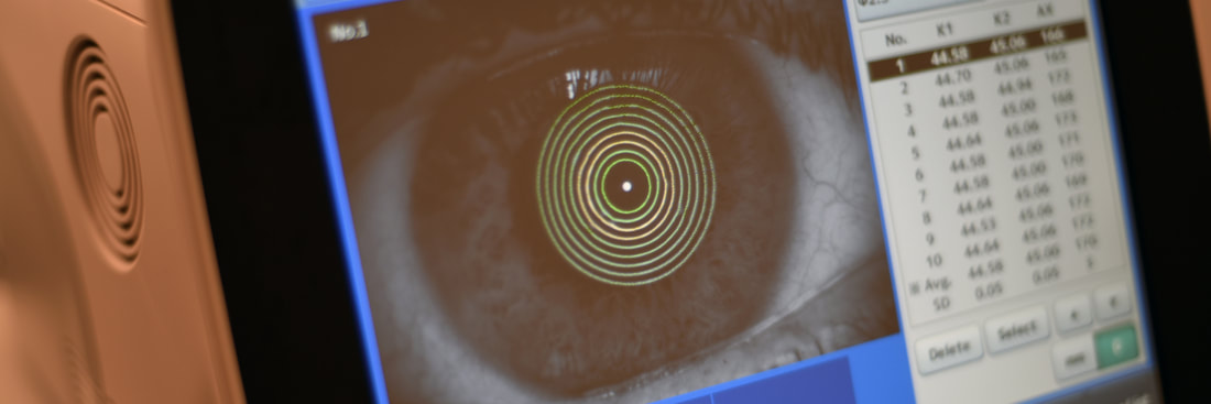
Keratoconus is diagnosed on clinical examination and by using advanced cornea tomography scans such as the Pentacam scans, available at IRIS surgery. These scans are important as they pick up early disease and serial scans will pick up any progression of keratoconus. It is important to have regular scans as CXL should be performed when progression is noted. There are situations when the disease remains stable and can be conservatively monitored without any surgery.
Please view above (first 2) embedded videos for further information on Keratoconus.
Fuchs' Endothelial Dystrophy
What is Fuchs' Endothelial Dystrophy (FED)? What are the symptoms?
FED is a disease of the cornea, specifically of the cells that line the inner layer of the cornea. These cells pump fluid out of the cornea to keep the cornea clear and transparent. When they are unhealthy, the fluid in the cornea builds up and it becomes hazy, resulting in poor vision. Typically, in the early stages, vision is hazy in the morning and it clears up as the day progresses because fluid in the cornea evaporates away when the eyes are open during the day. In the late stages, vision remains blurry all the time as there is too much fluid for evaporation to compensate. In the advanced stages, blisters can form on the cornea and these can break and cause significant eye pain.
How is FED diagnosed? How is it treated?
Fuchs' endothelial dystrophy can be diagnosed on examination. A few additional scans to assess the health of your cornea are usually done. There is no cure for Fuchs' but there are effective treatments which can maintain your vision depending on the stage of your disease. In general, cornea transplant surgery is required only when your vision is affected.
With new techniques in cornea transplantation, selective transplantation of only the diseased layer of the innermost cells are exchanged in a process called endothelial keratoplasty. There are 2 types of endothelial keratoplasty:
What is a Cornea Transplant?
Cornea for transplantation comes from a person who has elected to donate their organs. They are precious and should be treated with respect. I ask that persons who are fortunate enough to receive a donation to improve their vision look after their transplants and be compliant with long term eye drops. A long term commitment to follow-up will be required to ensure the health of their transplant.
Contact us
Keratoconus
What is Keratoconus?
The cornea is the clear transparent window at the front of your eye in the shape of a round dome. If you wear contact lenses, the cornea is where the contact lens rests on. It is responsible for accurately focusing light into the back of the eye. In keratoconus, the cornea thins out and becomes uneven, resulting in an irregular cone shaped cornea. As a result, the light rays entering the eye become out of focus and your vision becomes blurry and distorted.
What are the symptoms of Keratoconus? How is keratoconus treated? What is Cornea Cross Linking (CXL)?
Keratoconus starts in your teens. Early symptoms are worsening vision that require regular change in glasses with increasing astigmatism. Once keratoconus progresses to the late stages, your vision will be poor and may not be correctable with glasses or contact lenses. When this happens a cornea transplant is required (CAIRS, DALK or PK). Cornea Cross Linking (CXL) is a new treatment which can halt or slow the progression of keratoconus. This aims to prevent the late stages of disease. However this is only effective in the stage of early progressive disease. This is why early diagnosis and monitoring in keratoconus is important.
How is Keratoconus diagnosed? How do I know if I have early progressive disease?
Keratoconus is diagnosed on clinical examination and by using advanced cornea tomography scans such as the Pentacam scans, available at IRIS surgery. These scans are important as they pick up early disease and serial scans will pick up any progression of keratoconus. It is important to have regular scans as CXL should be performed when progression is noted. There are situations when the disease remains stable and can be conservatively monitored without any surgery.
Please view above (first 2) embedded videos for further information on Keratoconus.
Fuchs' Endothelial Dystrophy
What is Fuchs' Endothelial Dystrophy (FED)? What are the symptoms?
FED is a disease of the cornea, specifically of the cells that line the inner layer of the cornea. These cells pump fluid out of the cornea to keep the cornea clear and transparent. When they are unhealthy, the fluid in the cornea builds up and it becomes hazy, resulting in poor vision. Typically, in the early stages, vision is hazy in the morning and it clears up as the day progresses because fluid in the cornea evaporates away when the eyes are open during the day. In the late stages, vision remains blurry all the time as there is too much fluid for evaporation to compensate. In the advanced stages, blisters can form on the cornea and these can break and cause significant eye pain.
How is FED diagnosed? How is it treated?
Fuchs' endothelial dystrophy can be diagnosed on examination. A few additional scans to assess the health of your cornea are usually done. There is no cure for Fuchs' but there are effective treatments which can maintain your vision depending on the stage of your disease. In general, cornea transplant surgery is required only when your vision is affected.
With new techniques in cornea transplantation, selective transplantation of only the diseased layer of the innermost cells are exchanged in a process called endothelial keratoplasty. There are 2 types of endothelial keratoplasty:
- Descemet's Membrane Endothelial Keratoplasty (DMEK). The thinnest possible corneal transplant measuring below 20 microns which give better visual results. This is the latest technique in endothelial keratoplasty where only the descemets' membrane and endothelial cells are transplanted. See her surgical video on a new DMEK insertion technique Dr Chong developed here.
- Descemet's Stripping Automated Endothelial Keratoplasty (DSAEK). Ultrathin layer of cornea is cut by an automated process and a thin layer of stroma along with descemet's membrane with it endothelial cells are transplanted. The DSAEK graft is thicker (90-180 microns) than DMEK and is an established treatment modality.
- Descemetorhexis Without Endothelial Keratoplasty (DWEK). This is an experimental technique where the sick endothelium is surgically removed, but no corneal transplant is performed, and the cornea is left to heal on its own. This is currently performed in a clinical trial setting if eligibility criteria is fulfilled. See here article written by A/Prof Chong for more information.
What is a Cornea Transplant?
Cornea for transplantation comes from a person who has elected to donate their organs. They are precious and should be treated with respect. I ask that persons who are fortunate enough to receive a donation to improve their vision look after their transplants and be compliant with long term eye drops. A long term commitment to follow-up will be required to ensure the health of their transplant.
Contact us